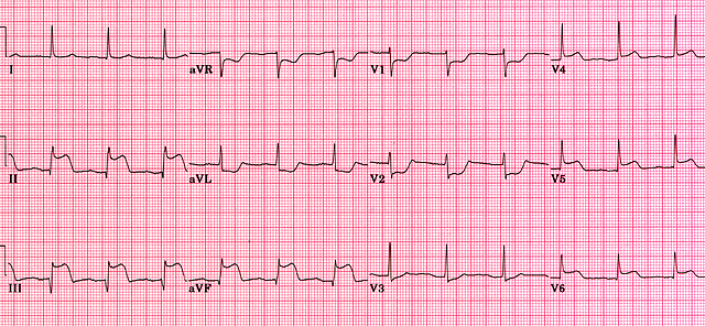
Acute inferior MI
Leads II, III and aVF reflect electrocardiogram changes associated with acute infarction of the inferior aspect of the heart. ST elevation, developing Q waves and T wave inversion may all be present depending on the timing of the ECG relative to the onset of myocardial infarction. Most frequently, inferior MI results from occlusion of the right coronary artery. Conduction abnormalities which may alert the physician to patients at risk include second degree AV block and complete heart block together with junctional escape beats. Note that the patient below is also suffering from a concurrent posterior wall infarction as eveidenced by ST depression in leads V1 and V2.
