Pulmonary embolism
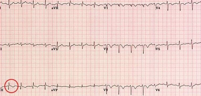
Electrocardiogram abnormalities can be observed in a minority of patients presenting with pulmonary embolism. These changes are rarely diagnostic unless greater than 50% of the pulmonary vascular compartment is occluded. Pulmonary embolism increases resistance to blood flow to the right side of the heart, commonly resulting in cor pulmonale involving right atrial enlargement and right ventricular dilation or hypertrophy. Lead III demonstrates ECG changes which mimic acute inferior myocardial infarction (circled below). These changes include an increase in the normal Q wave amplitude, minimal ST segment elevation, and often shallow T wave inversion. Pulmonary embolism is differentiated from acute inferior MI by the absence of these changes in the other inferior leads (II and aVF). Elevated ST segments, increased S wave amplitude and inverted T wave polarity may sometimes be seen in the precordial leads.