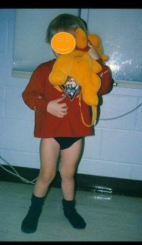
Deformity can be quantified and followed in both cases by clinical measurements of the femoral tibial axis and/or by the distance between the medial femoral condyles or medial malleoli when standing. In cases of physiological genu valgum or varum, reassurance is all that is needed. If the diagnosis is uncertain, then it is helpful to follow the patient clinically over time to determine whether there is progression or resolution of deformity. | | 3-year old boy with physiologic genu valgum. This requires no treatment. | |
|
|
Indications for x-rays are similar to those for genu varum. Deformity can be quantified and followed in both cases by clinical measurements of the femoral tibial axis and/or by the distance between the medial femoral condyles or medial malleoli when standing. In cases of physiological genu valgum or varum, reassurance is all that is needed. If the diagnosis is uncertain, then it is helpful to follow the patient clinically over time to determine whether there is progression or resolution of deformity. |