Queen's University MSK Unit Clinical Cases:
Upper limb pain and dysfunction
This module will enable you to apply your knowledge of appendicular anatomy to the analysis of common upper extremity musculoskeletal injuries. Please complete the quiz, review the references and consider the case scenarios in advance of the related session. In class, you will work in teams to explore these case scenarios in more detail. You may bring resources such as notes, texts and wireless-enabled laptops to help you work through and solve the problems posed to your team.
Objectives
By the end of this module and the related classroom session, the student will be able to:
Relevant MCC Objective(s): 67-1-2-2
Required preparation
Please read the following reference prior to the session:
You should also review relevant anatomy using the course text as well as Dr. Reifel's lectures and lab sessions related to the upper shoulder and elbow.
Optional preparation
Review the companion module on Examination of the Shoulder
(you will need to do this prior to your Expanded Clinical Skills sessions in Phase IIA)
The purpose of this quiz is to allow you to assess your knowledge of shoulder and elbow anatomy prior to the scheduled case-base session. The score will not contribute to your class grade however completion is mandatory. Please print out a blank copy of this quiz by clicking here (PDF - MEdTech Username and password required) and submit the completed quiz at the beginning of the related classroom session. (Alternatively, you can print the completed file from your browser once completed by clicking on the quiz, selecting print from the File menu and choosing "print only the selected frame".)
Question 1
Question 2
Question 3
Question 4
Question 5
Question 6
Question 7
Question 8
Question 9
Question 10
Student name: ______________________________
Case 1
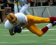
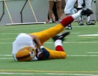
You are attending the annual Homecoming football game and witness the following injury. The player is helped off the field and does not return to play for the remainder of the game.
Image credit: D. Bardana
Later that day, you encounter the player as a patient in the Emergency Room where you are working. He is a right-handed fourth-year Engineering student who complains of pain in the right shoulder subsequent to this fall. He is reluctant to abduct or elevate the right arm as he reports that these motions cause pain and a "crunching" feeling. You notice an obvious deformity (see picture below). The patient is most tender just lateral and antero-inferior to the deformity.
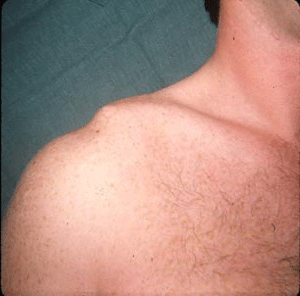
Image credit: L. Davidson
Questions for discussion:
Case 2
A 22-year old mechanic presents with intermittent right shoulder discomfort and 'weakness'. He has had symptoms for several years, worse when he works overhead. He reports feeling an uncomfortable popping sensation when his arm is elevated and externally rotated. He has given up playing baseball in a recreational league because of these symptoms. He does not report any specific history of injury, although admits that as a teenager he was involved in many different sports and had many visits to the Emergency room for a variety of sports-related injuries.
On examination, the patient appears health and muscular. There is no deformity and he has full range of motion of both shoulders in all directions. The physical examination maneuver pictured below reproduces his symptoms.
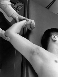
Image credit: D. Bardana
Questions for discussion:
Case 3
A 53-year old woman presents with pain and dysfunction of her right shoulder. She has had symptoms for the past three months and denies any injury. She feels that the problem is getting worse and is now having trouble with day-to-day activities such as washing her hair and reaching overhead. The pain is dull and constant and worse with attempted arm elevation or if she lies on the right side in bed at night. She has no other joint problems and is healthy other than non-insulin dependent diabetes that is currently well-controlled with diet and medication.
When you examine her, you notice a difference in the motion of the right and left shoulders. In particular, she lacks internal rotation and abduction of the right shoulder.
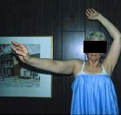
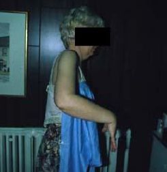
Image credit: G.M. Taylor
Questions for discussion:
Case 4
A 17-year old athlete requests an urgent appointment to see you because of bilateral lateral elbow pain (right greater than left). The pain has been present for three months and has coincided with volleyball season. He is very anxious to continue playing because his team is doing well this season and is likely to win the regional championships. In addition, he is aware that there are some university athletic scouts who are interested in his performance. He has applied ice after several games with some improvement and takes the occasional ibuprofen tablet if the pain is severe. He is otherwise healthy with no other active musculoskeletal issues.
On physical exam you notice that the patient has no deformity, redness or swelling about the elbows. He has full range of motion of both shoulders, elbows and wrists however resisted wrist extension on the right reproduces the elbow pain. He is locally tender over the lateral aspect of the elbow.
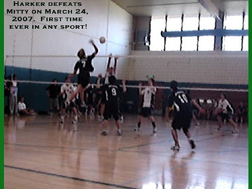
Image credit: milesgehm
Questions for discussion: