Queen's University MSK Unit Clinical Cases
Lower limb pain and dysfunction
This module will enable you to apply your knowledge of appendicular anatomy to the analysis of common lower extremity musculoskeletal injuries and pathologies. Please complete the quiz, review the references and consider the case scenarios in advance of the related session. In class, you will work in teams to explore these case scenarios in more detail. You may bring resources such as notes, texts and wireless-enabled laptops to help you work through and solve the problems posed to your team.
Objectives
By the end of this module and the related classroom session, the student will be able to:
1. Recognize common injuries and pathologies of the hip and knee,
2. Identify the likely anatomical source of pathology based on historical information and physical findings of hip and knee dysfunction,
3. Identify common radiologic abnormalities characteristic of hip and knee injuries and pathologies.
Relevant MCC Objective(s): 67-1-2-3
Required preparation
Please read the following reference prior to the session:
Lawrence, P.F. (2007) Essentials of Surgical Specialities, 3rd edition, p. 245-247 and 249-256 (focusing on the lower extremity topics)
You should also review relevant anatomy using the course text as well as Dr. Reifel's lectures and lab sessions related to the hip and knee.
1. Gluteal Region and Thigh
2. Leg and Knee
Test your knowledge of anatomy
The purpose of this quiz is to allow you to assess your knowledge of hip and knee anatomy prior to the scheduled case-base session. The score will not contribute to your class grade however completion is mandatory. Please print out a blank copy of this quiz from the MedTech page associated with this session and submit the completed quiz at the beginning of the related classroom session.
Question 1
Question 2
Question 3
Question 4
Question 5
Question 6
Question 7
Question 8
Question 9
Question 10
Case 1
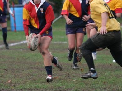
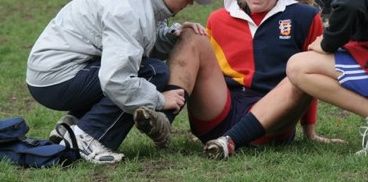
During the last moments of the final game in the inter-university women's rugby tournament, Queen's star player is tackled by a member of the opposing team. She suffers a medial blow to her right knee and falls to the ground. She is unable to weightbear and is assisted off the field by her trainer. The right knee becomes swollen immediately. Both flexion and extension of the joint are limited by a combination of swelling and pain.
Image credit: D. Bardana
Think about these questions in advance of the session:
Case 2
You are the clinical clerk working in the Emergency Department during the Friday afternoon shift. Mrs. McEwan is a 77-year old woman who presents with pain in her distal femur and medial knee. She came by ambulance as she was unable to cope alone at home. The pain began a week ago following a minor fall (she slipped on a loose rug) in her home. She has had difficulty ambulating ever since then. The resident that you are working with is busy assessing a patient in septic shock. Based on the triage nurse's note, he has ordered the x-ray pictured below and asked you to perform a full assessment of the patient.
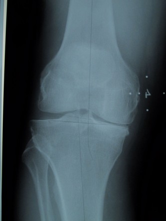
Image credit: M. Harrison
Case 3
Arun, a 36-year old graphic designer presents with a four-month history of right groin pain. He does not recall any injury. The pain is gradually increasing and he now has to use crutches to walk long distances. He has an obvious Trendelenburg limp when he walks into your office. This patient's past medical history is significant for asthma and eczema. He has been treated with a combination of oral inhalers (salbutamol and corticosteroids) and oral prednisone (once or twice a year when he has flare-ups) for the past 20 years.
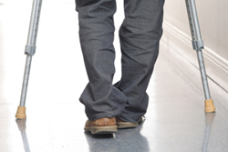
Image credit: unknown
Case 4
Jesse is a fifteen-year-old who presents with a six-month history of right knee swelling, restriction of motion and pain. He does not recall any specific injury, although he is generally very active in sports (hockey, volleyball, basketball) and has had many bangs and bruises over the last several years. His problem has been slowly progressive over the last few months and has caused him to drop out of gym and not participate in hockey this year. He is otherwise healthy and has no allergies. His only medications are occasional over-the-counter analgesics (acetaminophen and ibuprofen) which he takes rarely (once or twice a week) if his knee is particularly sore.
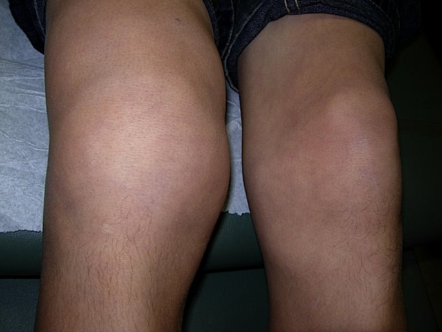
Image credit: L. Davidson